CT Imaging of the heart has been available since the introduction of 64-slice scanners in 2005/2006, and was improved with later scanners that had higher speeds and greater volume coverage. The technique is non-invasive, requires no hospitalization and is easily tolerated by most patients. The newer scanners and techniques require only modest doses of radiation and contrast agent. The scan provides a high-resolution anatomic image of the heart and of the coronary arteries.
While CT imaging of the heart is a powerful tool for diagnostics and supporting therapeutics of different heart diseases and conditions, the main application so far is triage of Coronary Heart Disease (CHD) prior to Invasive Coronary Angiography in a catheterization laboratory. As discussed above, a large number of invasive catheterizations end up not finding substantial obstruction. These unnecessary interventions can be avoided by prior imaging of the coronaries with CT. In addition, CT cardiac imaging can be an effective tool for identifying patients that need non-invasive medical treatment to reduce cardiovascular risk as well as facilitating early treatment for patients that need immediate intervention.
Among the numerous clinical studies supporting the effectiveness of cardiac CT it is worth mentioning the SCOT-HEART study (M. C. Williams et. al, JACC Vol. 67, No. 15, 2016: 1759–68). 4,146 patients with suspected angina were randomly assigned to standard care vs. standard care plus Coronary Computed Tomography Angiography (CCTA). While there were similar rates of referral to invasive angiography in the two groups, in those allocated to CCTA the invasive angiography was less likely to demonstrate normal coronary arteries (20 vs. 56) and more likely to show obstructive CHD (283 vs. 230). More preventive therapies (283 vs. 74) were initiated after CCTA and fatal and nonfatal myocardial infarction was almost halved (!) in patients undergoing CCTA compared with those assigned to standard care (26 vs. 42 after 1.7 years follow up). In other words, in this study CCTA was demonstrated to actually save lives. A five years follow up on the SCOT-HEART study was published in 2018 (D. E. Newby et. al. NEJM.org August 25, 2018). The trends observed at three years persist. In particular, death from coronary heart disease or nonfatal myocardial infarction at median duration of 4.8 years was lower in the CCTA group than in the standard-care group (2.3% [48 patients] vs. 3.9% [81 patients].
Following the SCOT-HEART trial and other evidence, in its Clinical Guidelines 95 (2016) the UK National Institute for Health and Care Excellences (NICE) has recommended cardiac CT as the first-line investigation for all patients presenting with chest pain due to suspected coronary artery disease.
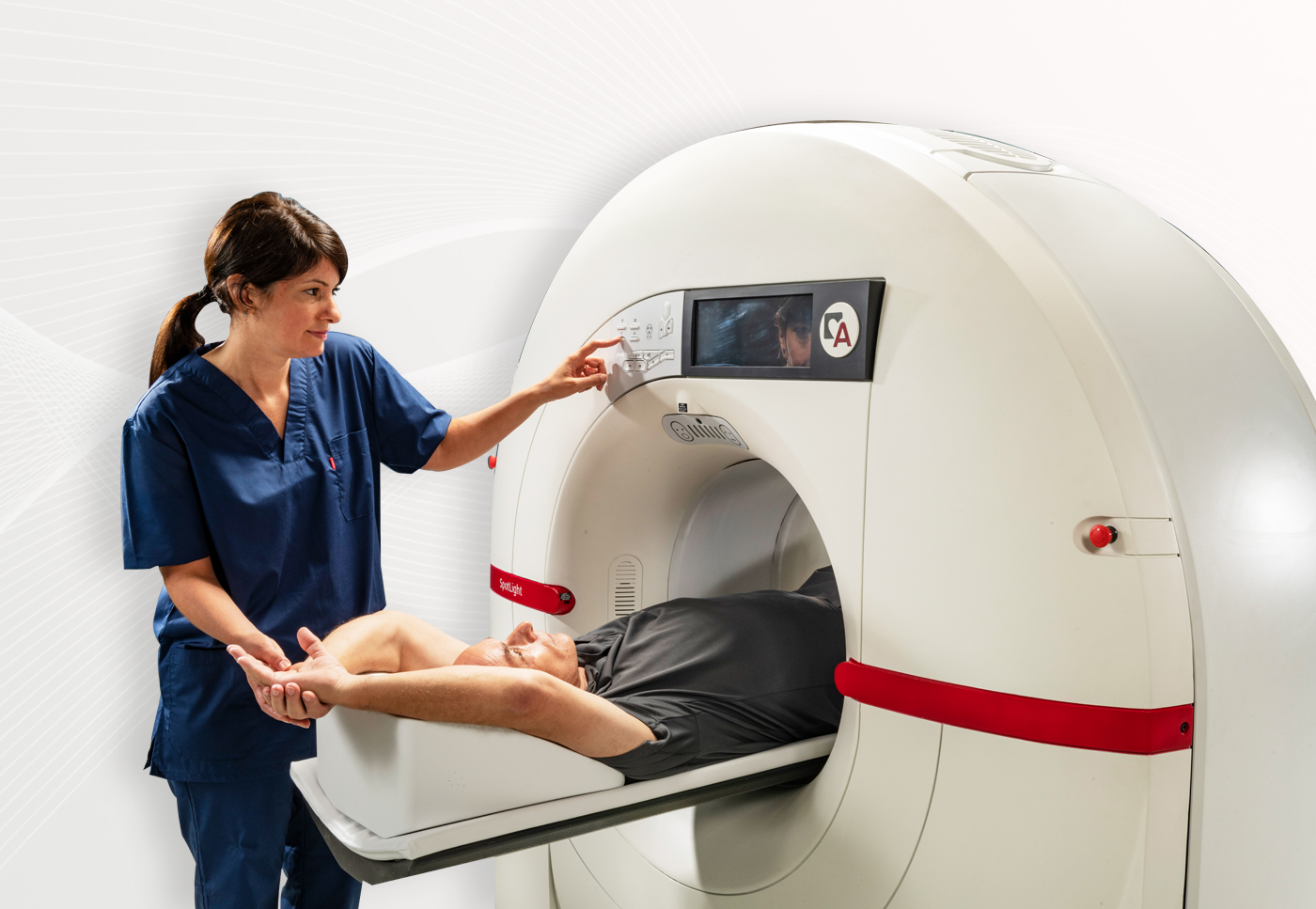
Until the introduction of CardioGraphe by Arineta and GE, there were no dedicated cardiac CT scanners on the market. The procedures are typically done on whole body, multi-purpose radiology scanners, with compromised results when used for cardiac imaging tasks. The CardioGraphe clinical results have shown that a whole heart, single-beat, special-purpose cardiovascular scanner greatly improves clinical outcomes.