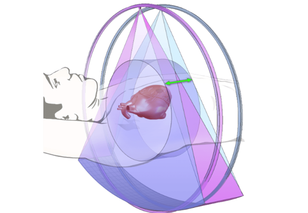
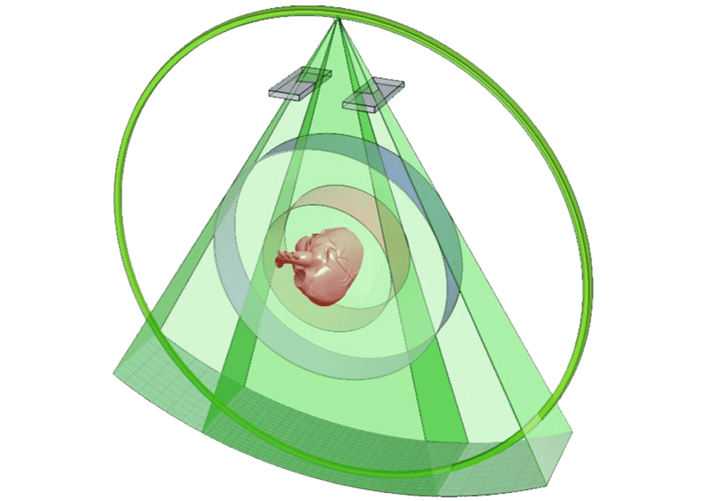
Technology
Stereo CT® Technology
Unlike conventional single- and dual-source CT scanners, CardioGraphe uses dual overlapping cone beams to achieve 140 mm coverage. Stereoscopic imaging provides a more complete data set than mono-beam CT, for better, artifact-free images. Stereoscopic imaging also optimizes the scanner’s dose utilization.
Read More
Key elements of the technology are:
- Dual, specially designed x-ray tubes
- High-voltage generator capable of ultra-fast x-ray beam switching between the two sources
- CardioGraphe detector for low-noise, high-rate acquisition
- Unique Stereo CT reconstruction algorithm for optimal integration of data from the two sources into a single image set

Focused Field-of-view
CardioGraphe uses Focused Field-of-View technology for high quality imaging of an area-of-interest, while reducing the radiation dose in peripheral anatomies outside the area-of-interest (shown for a single x-ray source).
Radiation outside the field-of-view (user-selected 250 mm or 160 mm diameter) is attenuated by a factor of 10 – 20 (depending on kVp). (Actual dose saving depends on a combination of kVp, selected FOV diameter, and patient size.)
